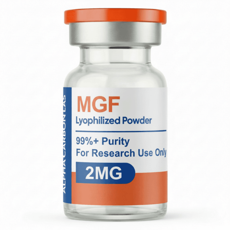
2MG
This product is for research purposes only. Not for human consumption.
Purity: >98% (HPLC verified)
Formulation: Lyophilized powder
Molecular Formula: C37H51N9O10S
Molecular Weight: 813.93 g/mol
CAS Number: 12020-86-9
PubChem CID: N/A
MGF
Overview
Mechano Growth Factor (MGF), also known as IGF-1Ec or by its full designation as Insulin-like Growth Factor-1 Splice Variant Ec, is a locally-acting splice variant of the IGF-1 gene that is produced specifically in muscle tissue in response to mechanical loading, exercise-induced muscle damage, or stretch stimuli - earning its name from this mechano-sensitive expression pattern that distinguishes it from the systemically-circulating, liver-derived IGF-1 (often called systemic IGF-1 or IGF-1Ea) that is regulated primarily by growth hormone. The discovery of MGF emerged from research by Geoffrey Goldspink and colleagues in the 1990s-2000s investigating how muscles respond and adapt to mechanical stress at the molecular level. This work revealed that the IGF-1 gene, through alternative splicing of its mRNA, can produce multiple distinct IGF-1 isoforms with different C-terminal E domain sequences, each having unique biological properties. MGF contains a unique 49-52 amino acid E peptide domain (the exact length varies slightly depending on species and specific splice variant) that confers specific biological activities particularly relevant to muscle regeneration and hypertrophy. While systemic IGF-1 promotes general tissue growth, survival, and metabolism throughout the body, MGF acts predominantly in a paracrine/autocrine manner - being produced locally within skeletal muscle fibers in response to mechanical signals and acting on nearby cells within the same muscle microenvironment. The mechanical stimuli that trigger MGF expression include the stretching forces during eccentric muscle contractions (lengthening under load, such as lowering a weight), the cellular damage and membrane disruption from intense exercise, and the inflammatory signaling that accompanies muscle injury. Following resistance exercise or muscle injury, MGF expression in the affected muscles increases dramatically within hours, reaching peak levels before declining over the following days as repair processes proceed. This temporal expression pattern positions MGF as an early-response factor in the muscle adaptation and repair cascade. The primary biological function of MGF centers on satellite cell activation and recruitment. Satellite cells are muscle-specific stem cells that normally exist in a quiescent (dormant) state along muscle fiber surfaces, but when activated by signals like MGF, they re-enter the cell cycle, proliferate to expand their numbers, and can differentiate into myoblasts that fuse with existing muscle fibers to support hypertrophy or fuse together to create new muscle fibers to replace severely damaged ones. MGF appears to be particularly potent at activating these satellite cells, more so than systemic IGF-1, potentially due to its unique E domain sequence creating preferential signaling properties. Beyond satellite cell effects, MGF activates protein synthesis pathways within existing muscle fibers, promotes myofiber hypertrophy, provides anti-apoptotic survival signals to protect muscle cells from programmed death during metabolic stress, and may have additional roles in muscle metabolism and performance. However, MGF has a critical pharmacological limitation: its plasma half-life is extraordinarily short, estimated at only 5-7 minutes, due to rapid proteolytic degradation by enzymes in blood and tissues and rapid renal clearance. This extreme instability means that systemically administered MGF is degraded almost immediately, achieving minimal systemic concentrations and limited distribution beyond the injection site. This pharmacokinetic challenge has led to two approaches in MGF research and application: local intramuscular injection directly into target muscles to achieve high local concentrations before degradation, or PEGylation (covalent attachment of polyethylene glycol chains) to create PEG-MGF with extended half-life, though the latter introduces its own complexities. MGF exists in a gray zone between established scientific research tool and unregulated research chemical, with substantial preclinical data demonstrating biological activity but limited controlled human studies, no regulatory approvals, and significant use in bodybuilding/athletic performance contexts based on extrapolation from mechanistic research rather than clinical efficacy data.
Mechanism of Action
MGF exerts its anabolic and regenerative effects primarily through activation of the IGF-1 receptor (IGF1R), a receptor tyrosine kinase that is expressed on muscle fibers, satellite cells, myoblasts, and numerous other cell types throughout the body. Despite being a splice variant of IGF-1, MGF binds to the same IGF1R that recognizes systemic IGF-1, though the unique C-terminal E domain of MGF may influence the kinetics, signaling output, or cellular context of receptor activation in ways that create functional distinctions from systemic IGF-1. When MGF binds to IGF1R on target cells, it triggers receptor dimerization and autophosphorylation of intracellular tyrosine kinase domains, creating docking sites for adaptor proteins including insulin receptor substrate-1 (IRS-1) and Shc. These adaptors initiate multiple downstream signaling cascades, with the two most important being the PI3K/Akt/mTOR pathway and the Ras/Raf/MEK/ERK (MAPK) pathway. The PI3K/Akt/mTOR axis is particularly crucial for MGF's anabolic effects: PI3-kinase activation generates PIP3 lipid second messengers that recruit and activate Akt kinase, which in turn activates the mechanistic target of rapamycin (mTOR) complex 1. mTORC1 is a master regulator of protein synthesis, and its activation leads to phosphorylation of downstream targets including p70S6 kinase and 4E-BP1, which ultimately increase ribosomal activity and translation of mRNA into proteins. This enhanced protein synthesis is essential for muscle fiber hypertrophy - the growth of individual muscle fibers through accumulation of contractile proteins including actin, myosin, and associated structural proteins. Akt also inhibits protein degradation pathways by phosphorylating and inactivating FoxO transcription factors that normally upregulate expression of atrophy-related genes including the E3 ubiquitin ligases MuRF1 and atrogin-1 (also called MAFbx), which tag muscle proteins for destruction by the proteasome. By simultaneously enhancing protein synthesis through mTOR and reducing protein degradation through FoxO inhibition, MGF shifts the net protein balance strongly toward anabolism. The MAPK pathway activated by MGF promotes cell proliferation and differentiation - critical processes for satellite cell function. Satellite cells are the key target cells for MGF's regenerative effects: in their normal quiescent state, satellite cells express low levels of myogenic regulatory factors and remain dormant, but MGF stimulation activates them to re-enter the cell cycle, undergo rounds of proliferation to expand their numbers (clonal expansion), and express myogenic determination factors including MyoD and myogenin that drive their differentiation into myoblasts and eventually fusion with existing muscle fibers. MGF appears to be more effective than systemic IGF-1 at this satellite cell activation function, possibly due to its unique E peptide influencing receptor signaling kinetics, duration, or subcellular localization, or through E peptide-mediated effects that are independent of IGF1R. Some research suggests the E peptide alone (separate from the receptor-binding IGF-1 core) may have biological activities, though this remains an area of active investigation. MGF signaling also provides survival signals through Akt-mediated inhibition of pro-apoptotic Bcl-2 family proteins, protecting muscle cells from programmed death during metabolic stress or injury. The localized production of MGF in mechanically-stressed muscle creates a microenvironment rich in this growth factor, exposing nearby satellite cells and muscle fibers to high concentrations that drive robust regenerative and hypertrophic responses.
Research Findings
Research on MGF has provided important insights into the molecular mechanisms of muscle adaptation to mechanical stress and injury, though the body of evidence consists primarily of preclinical studies with limited controlled human research. The foundational work by Goldspink's group characterized MGF as a distinct splice variant with unique expression patterns and biological properties. Studies using Northern blot, RT-PCR, and in situ hybridization demonstrated that MGF mRNA expression increases dramatically in skeletal muscle following eccentric exercise, muscle stretch, or injury, with peak expression occurring within hours of the stimulus and declining over subsequent days as systemic IGF-1 expression increases later in the recovery process. This temporal pattern suggested MGF functions as an early response factor initiating repair, while systemic IGF-1 supports sustained anabolism during later recovery phases. Cell culture studies using isolated myoblasts and satellite cells have demonstrated that MGF application stimulates their proliferation more effectively than equivalent concentrations of systemic IGF-1, supporting MGF's reputation as a particularly potent satellite cell activator. These in vitro studies have explored dose-response relationships, showing increased DNA synthesis, cell cycle entry, and expression of myogenic markers in MGF-treated cells. Animal studies have provided evidence for MGF's in vivo biological activity. Local intramuscular injection of MGF peptide or plasmid DNA encoding MGF into rodent muscles has produced increased muscle mass, enhanced muscle fiber size, accelerated recovery from induced injury, and improved strength compared to control treatments. Studies in models of muscle wasting, including aging-related sarcopenia, cachexia, and experimentally-induced atrophy, have shown that MGF treatment can partially attenuate muscle loss, supporting its potential therapeutic relevance. However, the short half-life of MGF means most animal studies have required frequent local injections, sometimes multiple times daily, which is not practically translatable to human applications. Research comparing MGF to systemic IGF-1 has explored their differential effects, with findings suggesting MGF may provide more localized effects with less systemic exposure, potentially reducing risks of systemic IGF-1 overexposure while enhancing local muscle adaptation. Human research specifically examining exogenous MGF administration is remarkably limited. There are no published large-scale controlled clinical trials of MGF for any indication, and most human data consists of studies measuring endogenous MGF expression in response to exercise or physiological conditions rather than exogenous administration studies. Research in humans has confirmed that MGF expression increases in skeletal muscle following resistance exercise, with higher expression correlating with muscle hypertrophy responses in training studies, supporting its physiological relevance to exercise adaptation. Gene expression studies in older adults versus younger individuals have shown age-related differences in MGF responses to exercise, potentially contributing to the blunted hypertrophy seen with aging. The absence of pharmaceutical-grade MGF products, controlled clinical trials, and regulatory approvals means that MGF use in humans occurs primarily in unregulated research contexts or performance enhancement settings, based on extrapolation from mechanistic research rather than direct human efficacy data.
Research Applications
- Muscle growth and hypertrophy research
- Muscle injury repair and regeneration studies
- Satellite cell activation and proliferation research
- Exercise-induced adaptation and recovery studies
- Muscle wasting and sarcopenia research
- Athletic performance and recovery research
- IGF-1 splice variant biology studies
- Localized anabolic signaling research
- Age-related muscle loss (sarcopenia) studies
- Cachexia and muscle wasting disorder research
Safety Profile
The safety profile of MGF in humans is poorly characterized due to the absence of controlled clinical trials, limited human research, and its status as an unregulated research chemical rather than a pharmaceutical therapeutic. Available safety information is primarily theoretical, extrapolated from IGF-1 biology, derived from limited animal studies, and based on anecdotal reports from non-clinical use contexts. The extreme short half-life of native MGF (5-7 minutes) means that systemically administered MGF would have very brief exposure, potentially limiting systemic toxicity risks but also limiting systemic efficacy unless administered locally or with extreme frequency. Local intramuscular injection, the primary route used in animal research and proposed for human use, theoretically confines MGF effects to the injected muscle and immediate vicinity before degradation, potentially reducing systemic exposure compared to long-acting systemic growth factors. However, even localized administration could produce tissue-level effects that warrant consideration. Theoretical safety concerns based on MGF's mechanism include: excessive IGF-1 receptor stimulation could promote uncontrolled cellular proliferation, particularly concerning given IGF-1's known role in promoting cell growth and potential contributions to cancer development and progression when chronically elevated - while short-lived local MGF pulses may differ from sustained systemic IGF-1 elevation in their cancer risk profile, this has not been adequately studied. Effects on satellite cell activation and muscle growth, while desirable for hypertrophy and recovery, could theoretically contribute to muscle imbalances, altered biomechanics, or dysregulated growth if not properly controlled. Hypoglycemia (dangerously low blood sugar) is a theoretical concern with any IGF-1 receptor agonist due to IGF-1's insulin-like effects on glucose uptake, though the brief duration of MGF action may limit this risk compared to longer-acting IGF analogs. The lack of pharmaceutical-grade MGF products means purity, identity, concentration, sterility, and absence of contaminants cannot be assured when obtaining MGF from research chemical suppliers - concerns about product quality, mislabeling, underdosing, overdosing, bacterial contamination, or presence of related but distinct peptides create significant safety unknowns. Injection-related risks including pain, inflammation, abscess formation, or systemic infection apply to any injectable compound, particularly when obtained outside pharmaceutical channels and potentially prepared or administered under non-sterile conditions. Long-term safety is completely uncharacterized - chronic use effects, potential for accumulation of effects even with short-acting peptides, impacts on normal IGF-1 homeostasis, and any delayed or cumulative toxicities are unknown. Potential for antibody formation against exogenous MGF with repeated exposure has not been evaluated, though the similarity to endogenous MGF might reduce immunogenicity risk compared to completely foreign peptides. Use in specific populations including adolescents (where effects on normal growth and development are concerning), individuals with existing cancers or precancerous lesions, those with diabetes or glucose metabolism disorders, and those with cardiovascular or other significant health conditions has not been studied and could pose particular risks. The absence of regulatory oversight, medical supervision in most use contexts, and established dosing guidelines means individuals using MGF are essentially conducting uncontrolled self-experimentation with unknown risk-benefit profiles.
Scientific References
Research Use Only
This product is intended for research purposes only and is not for human consumption, therapeutic use, or diagnostic applications. Please ensure compliance with all applicable regulations and institutional guidelines.